Benson, Aslanidi, Zhang, Holden, 2008
Model Status
This CellML model represents the ventricular epicardial cell. The model has been checked in both COR and PCEnv and it runs to recreate the published results. The units have been checked and they balance. As with the CellML version of the Hund and Rudy 2004 model, on which this particular model is based, a stimulus protocol has been added to allow the model to simulate action potentials for 5 seconds. The parameter 'tissue' has been added to switch between the original (default value 0) and 'tissue' (any other value, for example 1) models.
Model Structure
ABSTRACT: We have constructed computational models of canine ventricular cells and tissues, ultimately combining detailed tissue architecture and heterogeneous transmural electrophysiology. The heterogeneity is introduced by modifying the Hund-Rudy canine cell model in order to reproduce experimentally reported electrophysiological properties of endocardial, midmyocardial (M) and epicardial cells. These models are validated against experimental data for individual ionic current and action potential characteristics, and their rate dependencies. 1D and 3D heterogeneous virtual tissues are constructed, with detailed tissue architecture (anisotropy and orthotropy, due to fibre orientation and sheet structure) of the left ventricular wall wedge extracted from a diffusion tensor imaging data set. The models are used to study the effects of tissue heterogeneity and class III drugs on transmural propagation and tissue vulnerability to re-entry. We have determined relationships between the transmural dispersion of action potential duration (APD) and the vulnerable window in the 1D virtual ventricular wall, and demonstrated how changes in the transmural heterogeneity, and hence tissue vulnerability, can lead to generation of re-entry in the 3D ventricular wedge. Two class III drugs with opposite qualitative effects on transmural APD heterogeneity are considered: d-sotalol that increases transmural APD dispersion, and amiodarone that decreases it. Simulations with the 1D virtual ventricular wall show that under d-sotalol conditions the vulnerable window is substantially wider compared to amiodarone conditions, primarily in the epicardial region where unidirectional conduction block persists until the adjacent M cells are fully repolarised. Further simulations with the 3D ventricular wedge have shown that ectopic stimulation of the epicardial region results in generation of sustained re-entry under d-sotalol conditions, but not under amiodarone conditions or in control. Again, APD increase in M cells was identified as the major contributor to tissue vulnerability--re-entry was initiated primarily due to ectopic excitation propagating around the unidirectional conduction block in the M cell region. This suggests an electrophysiological mechanism for the anti- and proarrhythmic effects of the class III drugs: the relative safety of amiodarone in comparison to d-sotalol can be explained by relatively low transmural APD dispersion, and hence, a narrow vulnerable window and low probability of re-entry in the tissue.
 |
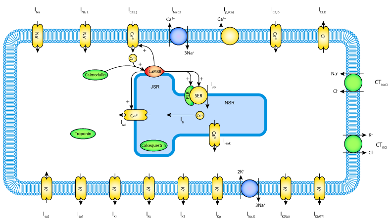
| Schematic diagram of the Hund and Rudy 2004 canine ventricular cell model (some of the model parameters and I_to equations have been updated in the current Benson et al. 2008 model). |
The original paper reference is cited below:
The canine virtual ventricular wall: a platform for dissecting pharmacological effects on propagation and arrhythmogenesis, Alan P. Benson, Oleg V. Aslanidi, Henggui Zhang and Arun V. Holden, 2008 Progress in Biophysics and Molecular Biology, 96, 187-208. PubMed ID: 17915298
